
Seminar Incucyte Zoom by A. Respa at MTZ - Kinetic live cell imaging inside your incubator
The system provides insight into active biological processes in real-time which is not possible using single-point and endpoint measures alone. The system resides within the controlled environment of a standard cell incubator. All imaging is completely noninvasive and non-perturbing to cell health. The system can process multiple plates, flasks and dishes in parallel and does not depend on shuttling plates into and out of the incubator.
The IncuCyte ZOOM® System allows you to:
- Complete long-term time-course experiments and then select optimal time points for post capture analysis.
- Follow the sequence of biological events, and then “rewind and replay” the experiment to understand and study the dynamics of what really happened to your cells while you were away.
- Build the data volume you need to validate and publish new ideas faster and more frequently.
- Perform routine cell health and proliferation studies alongside more advanced applications such as chemotaxis, T cell killing and stem cell differentiation.
- Use your time more efficiently. You can simply walk away and analyze your data remotely from any computer on the network.
Applications:
- Proliferation (confluence and cell counts)
- Cell Tracking / Cell Clustering Dynamics
- Apoptosis (caspase 3/7 for live-cell imaging)
- Cytotoxicity
- Dilution cloning (whole-well imaging)
- Chemotaxis
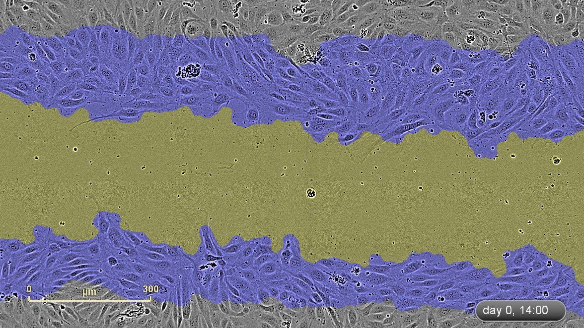
- Migration / Invasion / Wound Scratch
- Stem Cell monitoring and reprogramming
- 3D-Spheroids
- Phagocytosis
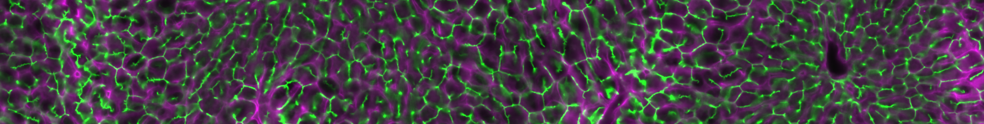
- Angiogenesis
- Neurite outgrowth and dynamics
- Reporter gene expression
- Viral studies
- Immune response – T-cell killing
- ROS / Hypoxia studies
Key Features:
- Operates from within a standard tissue culture incubator around the clock.
- Supports HD phase-contrast, green and red fluorescence imaging modes as well as whole well imaging.
- Automatically acquires and analyzes up to 2,000 images per hour.
- Supports over 300 different standard plates (including 96- and 384-well), T-flasks, dishes and microslides.
- Scans cell plates or tissue culture flasks according to a user defined schedule.
- Unlimited software licenses enable system control and data access at your convenience from any computer on your local network.